研究目的
To test and validate a recently designed and developed hand-held continuous-wave radio-frequency modulated diffuse optical spectroscopy probe in a clinical trial for breast cancer assessment.
研究成果
The RF-DOS probe can differentiate between malignant and healthy breast tissue in vivo with a sensitivity of 92 percent. The results are consistent with higher vascularization in cancerous tissue. Future work includes making the device more compact and integrating real-time data analysis.
研究不足
The probe did not detect the tumor in one case due to its small size and depth. The current hardware is bulky and not user-friendly, with data analysis and coefficient extraction performed separately.
1:Experimental Design and Method Selection:
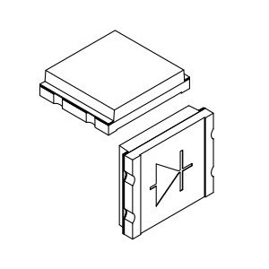
The study employed a hand-held continuous-wave radio-frequency modulated diffuse optical spectroscopy (RF-DOS) probe to measure optical properties of breast tissue in vivo. The probe uses an encapsulated LED light source emitting four wavelengths in the near-infrared range and two photodiodes for detection.
2:Sample Selection and Data Sources:
Fourteen patients diagnosed with breast cancer were recruited for the study. Measurements were taken on both the cancerous and contralateral healthy breast tissue.
3:List of Experimental Equipment and Materials:
The RF-DOS probe with an encapsulated LED (Marubeni America Corporation L690/750/800/850), two PIN photodiodes (Vishay Semiconductor TEMD5010X01), and an impedance spectroscope (Zurich Instruments Inc.-HF2IS) were used.
4:Experimental Procedures and Operational Workflow:
The probe was placed on the tumor location and the contralateral healthy site. Measurements were performed five times to negate the effects of body movement.
5:Data Analysis Methods:
The direct approach method was used to extract the concentration of deoxy-hemoglobin and oxy-hemoglobin from the measured data.
独家科研数据包��,助您复现前沿成果�����,加速创新突破
获取完整内容