研究目的
To develop a quantum dot-based fluorescence lifetime imaging microscopy (FLIM) glucose nanosensor for intracellular glucose detection, leveraging the unique optical properties of quantum dots (QDs) and the binding capacity of boronic acid to glucose.
研究成果
The QD-APBA conjugates developed in this study serve as effective glucose nanosensors, with the FLIM methodology providing significant advantages in sensitivity and selectivity for intracellular glucose detection. The long PL lifetimes of QDs allow for clear discrimination from cellular autofluorescence, enhancing the signal-to-background ratio. This approach holds potential for applications in cancer diagnosis by monitoring intracellular glucose levels.
研究不足
The study is limited by the pH dependency of the boronic acid–glucose interaction, requiring alkaline conditions for optimal sensitivity. Additionally, the selectivity towards glucose over other monosaccharides like fructose is not absolute, though physiological concentrations of interfering sugars are below the detection limit.
1:Experimental Design and Method Selection:
The study utilized CdSe/ZnS QDs modified with aminophenylboronic acid (APBA) to create QD-APBA conjugates for glucose sensing. The methodology included time-resolved spectroscopy and FLIM techniques to overcome limitations of PL intensity-based measurements.
2:Sample Selection and Data Sources:
MDA-MB-231 cells were used to test the intracellular detection of glucose. QD-APBA conjugates were synthesized and characterized for their response to glucose.
3:List of Experimental Equipment and Materials:
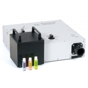
Instruments included a JASCO FP-6500 spectrofluorometer, FluoTime 200 fluorometer, MicroTime 200 fluorescence lifetime microscopy system, and a LIBRA 120 PLUS transmission electron microscope. Materials included CdSe/ZnS QDs, APBA, EDC, NHS, and various buffers.
4:Experimental Procedures and Operational Workflow:
QD-APBA conjugates were synthesized via EDC/NHS coupling reaction. The response to glucose was tested in solution and in MDA-MB-231 cells using FLIM. Cell viability assays were conducted to assess cytotoxicity.
5:Data Analysis Methods:
PL decay traces were analyzed using FluoFit 4.4 and SymphoTime 32 software. The PL average lifetime was calculated to quantify glucose levels.
独家科研数据包�����,助您复现前沿成果��,加速创新突破
获取完整内容